Dr Walker’s microscope
In our historic collection we have a microscope and glass slides belonging to Dr Walker. Dr Walker qualified in 1922 and was the first female doctor, looking after women’s health, at the psychiatric hospital Old Manor in Salisbury.
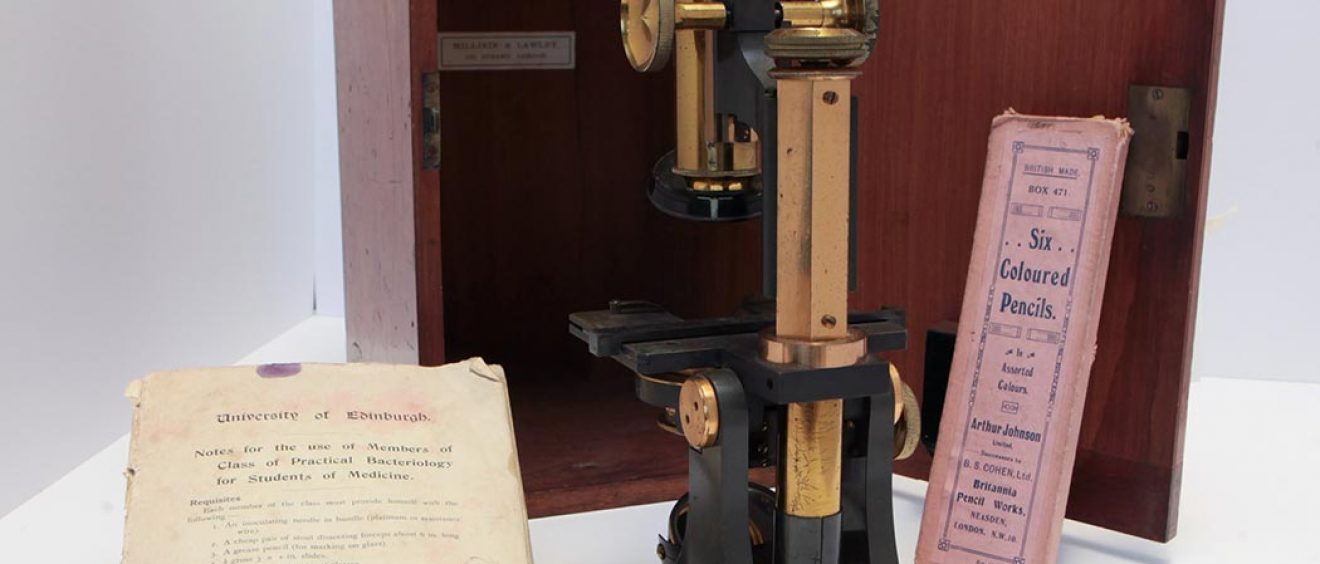
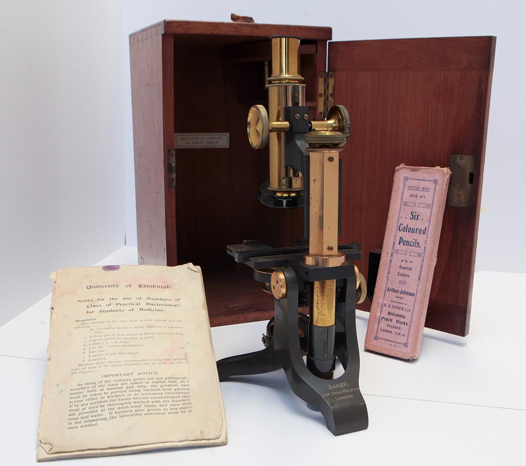
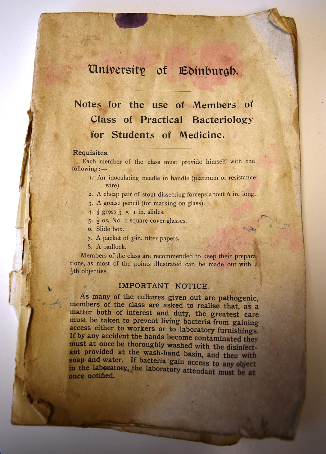
The images below show the brass and metal microscope, used in Dr Walker’s studies in the early 1920s, which is housed in a wooden case with key lock and leather strap handle. Inside the microscope box is a booklet from the University of Edinburgh, which gives students information about equipment required for Practical Bacteriology classes. Dr Walker has also written herself notes about the medical samples. Inside is also included a pack of coloured pencils, which were used to mark the glass slides. The cardboard slide box with drop down sides houses numerous prepared glass slides for pathology classes. Each one has been hand labelled with different medical conditions.


Did you know?
In the early 20th century there were very few women doctors. In 1881 there were only 25 in England & Wales. By 1911 there were 495. Shortages of medical staff during World War 1 meant there were greater opportunities for women in medicine and surgery.
How do microscopes work?
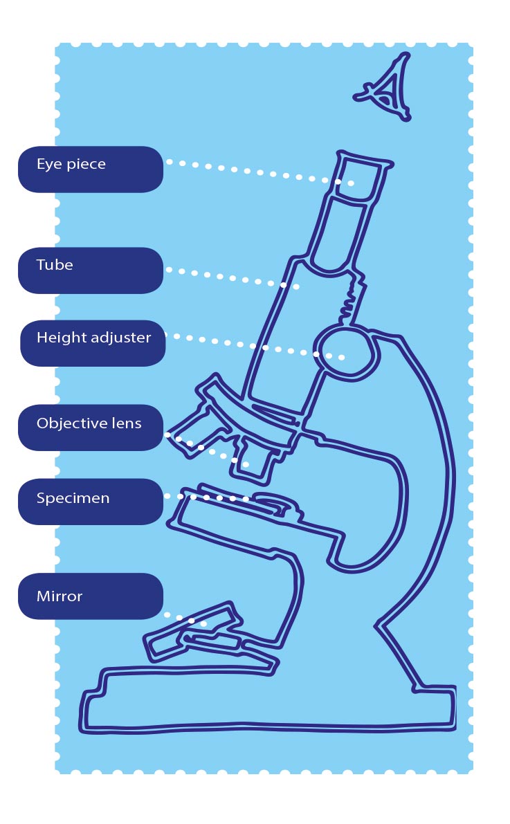
This optical microscope has two lenses. Lenses are curved pieces of glass that bend rays of light to make objects appear larger. Light from a mirror, at the base, is reflected up through the specimen, into the objective lens, which gives the first magnification. These lenses can be various sizes of magnification. On Dr Walker’s microscope there are 3 different strength objective lenses to choose from. The image is then increased again by the eyepiece lens, at the top, which is like a magnifying glass. You can see the enlarged image by looking into the eyepiece. The height adjuster helps to focus so you can see the object clearly. Our handy drawing below shows the different parts on an optical microscope.
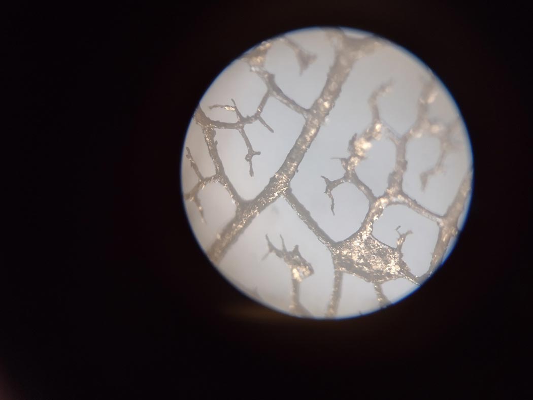
Looking through the microscope lens
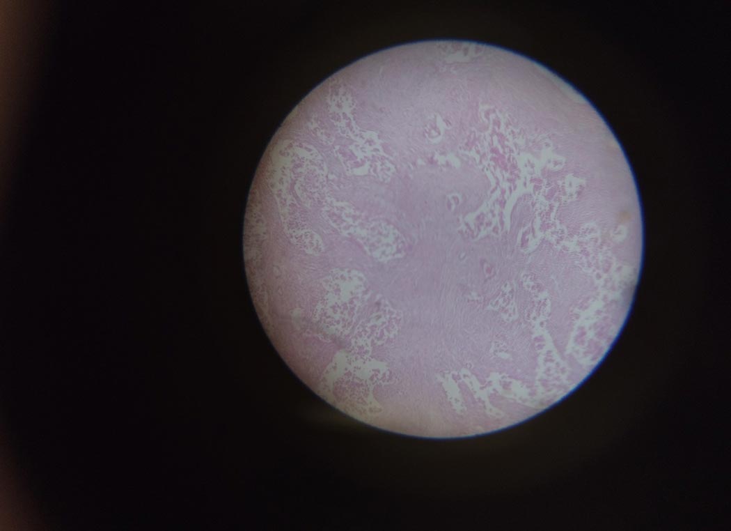
We used Dr Walker’s microscope to look at some of the historic samples. Below is a view of one which we photographed using a camera looking down the lens of Dr Walker’s microscope which still works today.
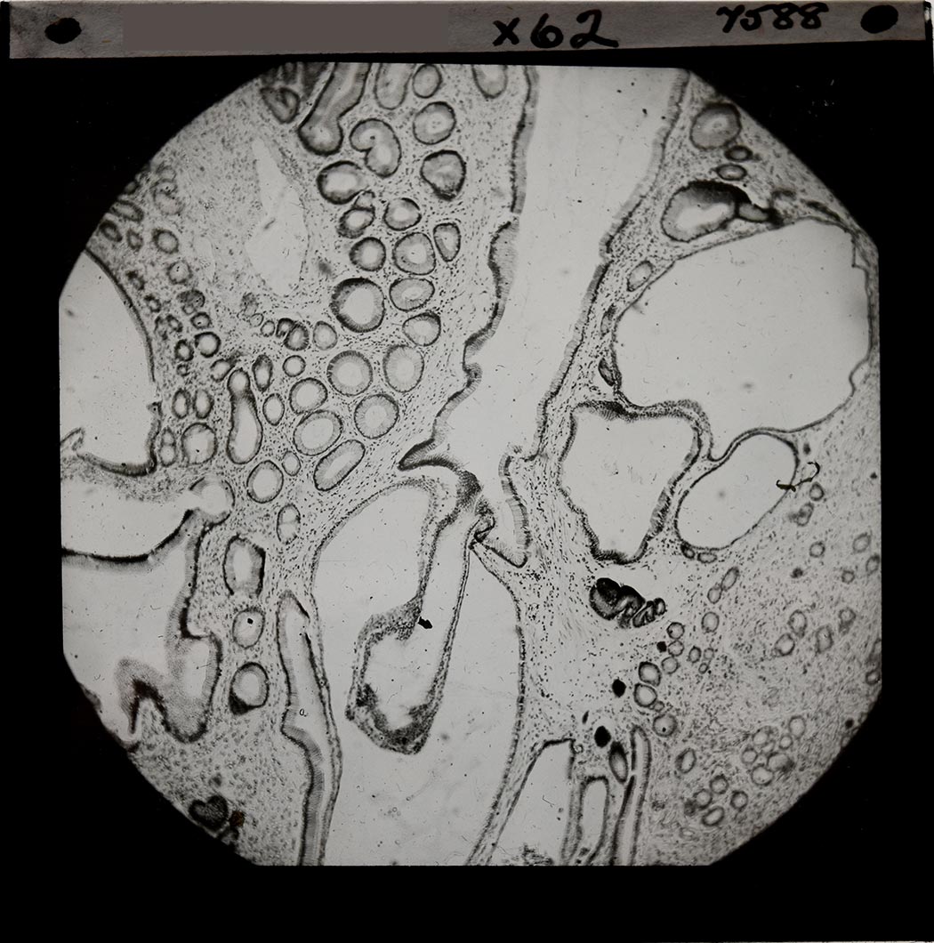
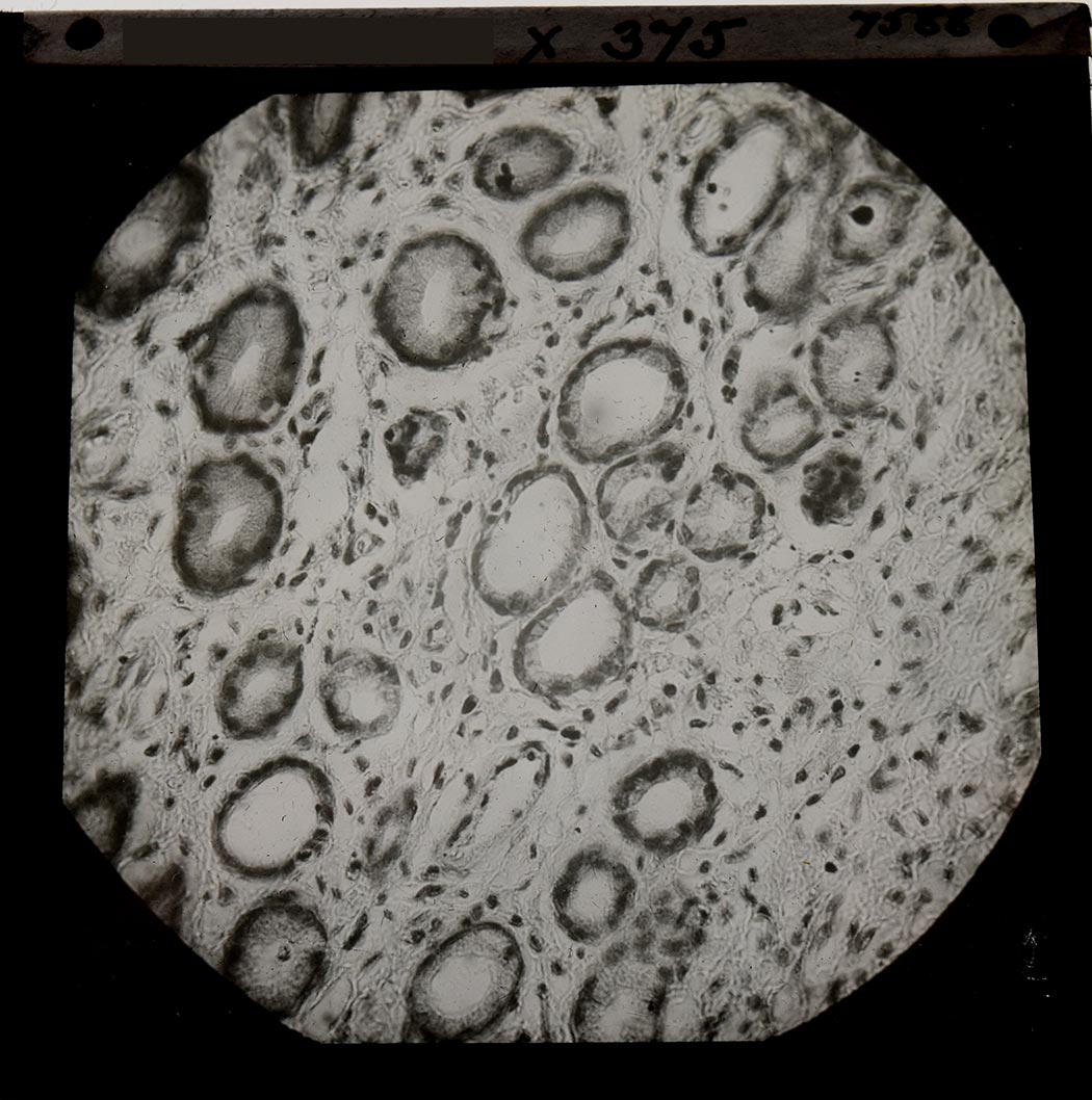
Also seen below are three small square glass plates from a Kodak box marked ‘Dr Roberts’ dating from early to mid 1950s. The first two show a section of tissue that has been removed and seen under a microscope magnification x 62. The third slide shows this sample at x 375 magnification.

Spots before your eyes!
We viewed newspaper print through Dr Walker’s microscope and photographed the image using a phone camera looking down the eyepiece. Below, you can clearly see the printing dots that make up colour images. This is where Cyan, Magenta, Yellow & Black (CMYK) dots make up different colours. By varying the size and density of each colour you can make up all the different shades you see in newspaper’s photographs. What do you think we are viewing through our microscope in the second image? HINT: the answer does grow on trees!