Marvellous MRIs
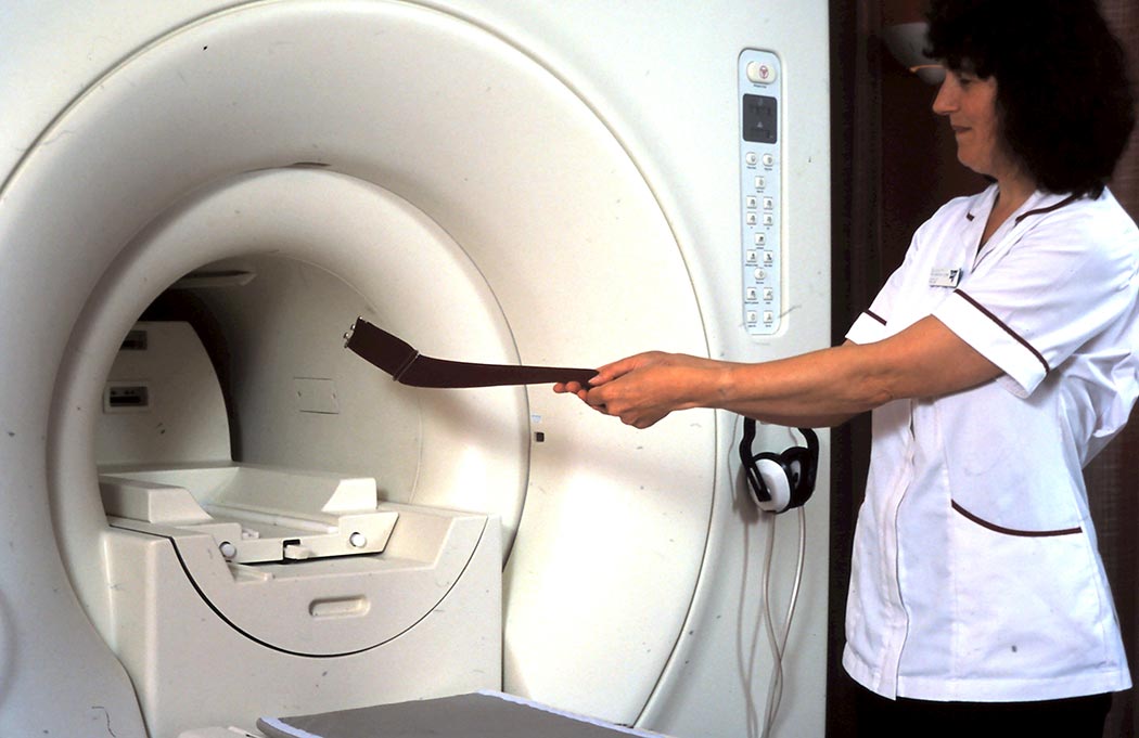
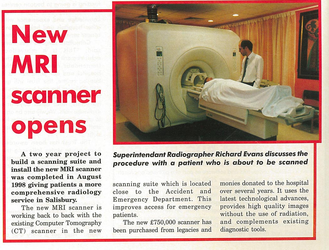
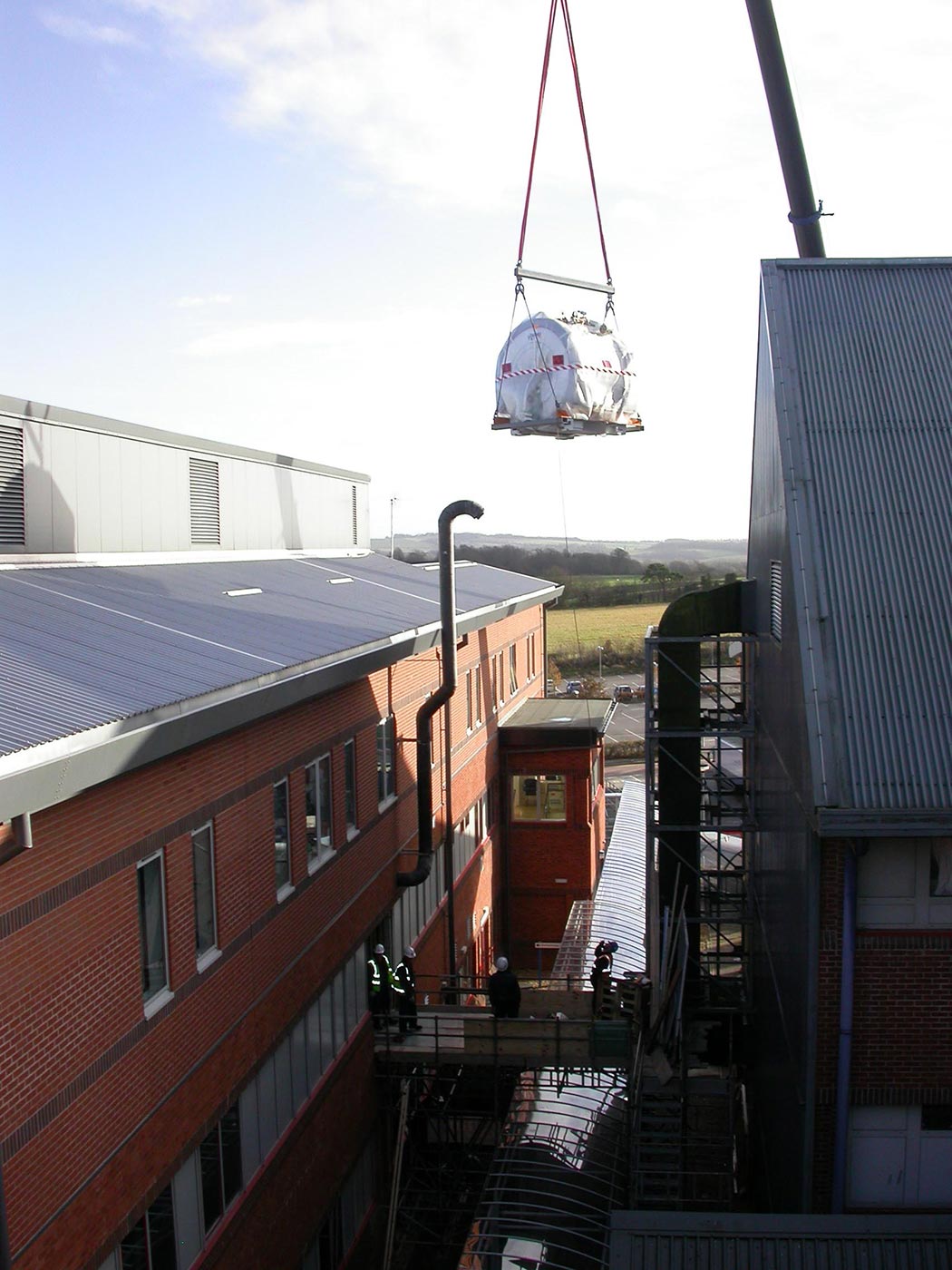
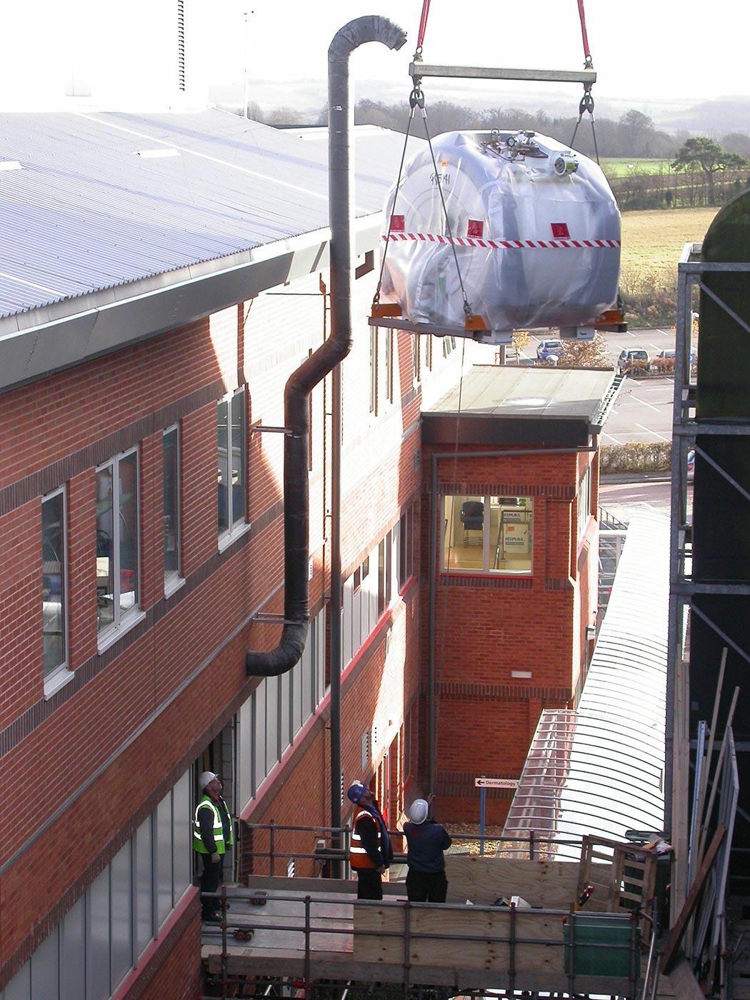
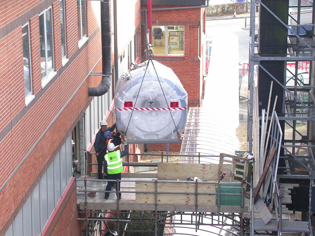
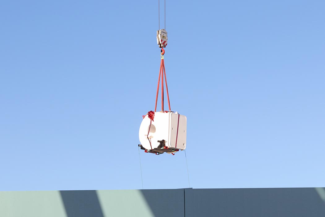
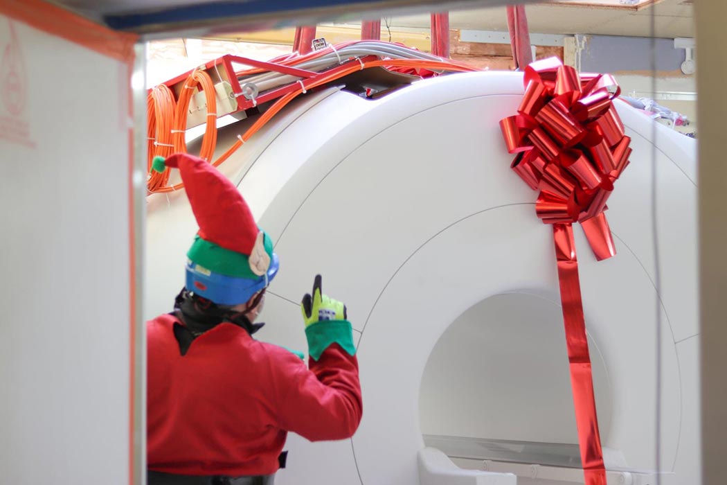
A look back through our archives has revealed some images taken in the early 1990s of Salisbury District Hospital’s MRI scanners. Depending on circumstances MRI scanners are generally replaced around every 10 years. A sequence of later images shows the delivery of a new MRI scanner being craned into the building. The design of the hospital includes this gap between the buildings to allow the crane to carefully lower the scanner. A removable wall allows the delivery and the heavy equipment can be moved directly into place. More recent images from December 2020, show an additional Stars Appeal funded MRI scanner being craned through the roof by Father Christmas and his elves. All this prompted us to take a closer look at how this technology works too.


How MRIs work
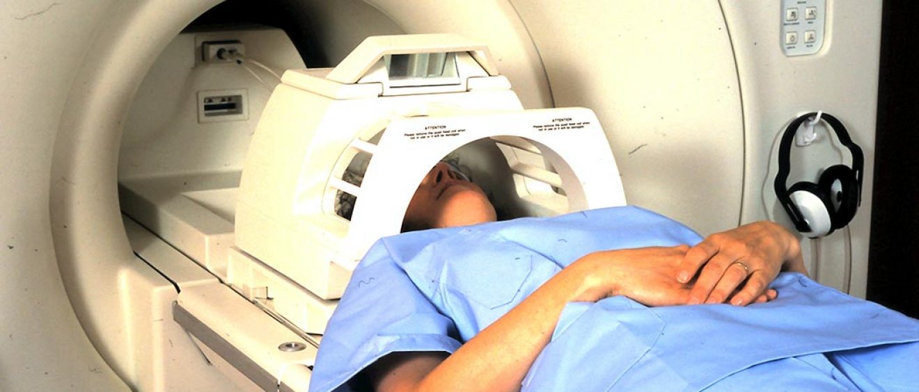
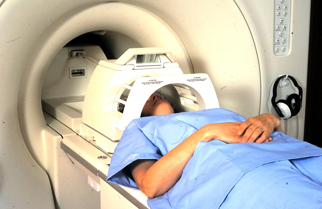
An MRI (magnetic resonance imaging) scanner is a large tube that contains powerful magnets. You lie inside the tube during the scan. At times the scanner will make loud tapping noises because the electric current in the coils is being turned on and off. It can take 15 to 90 minutes, depending on the size of the area being scanned and how many images are taken.
- The human body is mostly water, which is made of hydrogen and oxygen atoms. At the centre of each hydrogen atom is an even smaller particle called a proton. Protons are like tiny magnets. In the scanner, the protons in your body line up in the same direction like needle of a compass.
- Short bursts of radio waves knock the protons out of alignment and when the radio waves are turned off, the protons line up again.
- This sends out signals showing the location of protons and also helps to see various types of tissue because the protons in different structures line up at different speeds.
Read more about how MRI scans work at https://www.nhs.uk/conditions/mri-scan/
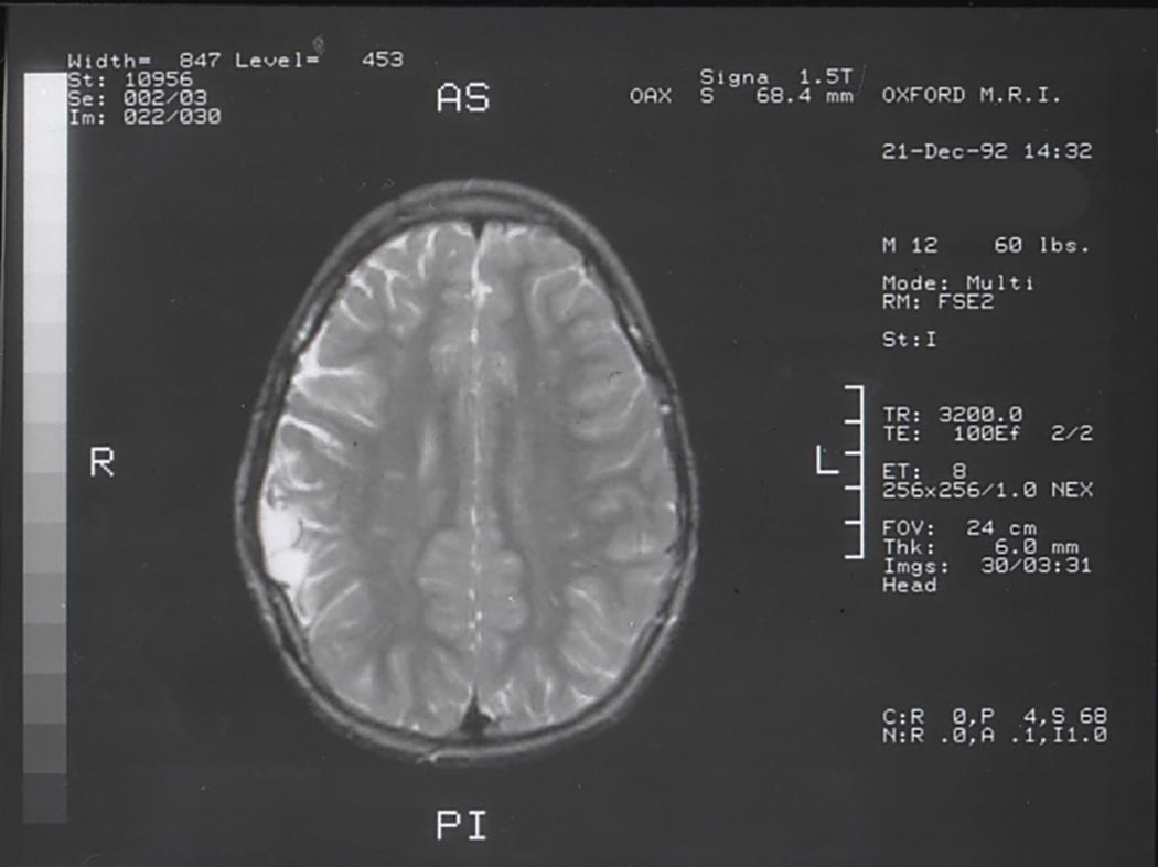
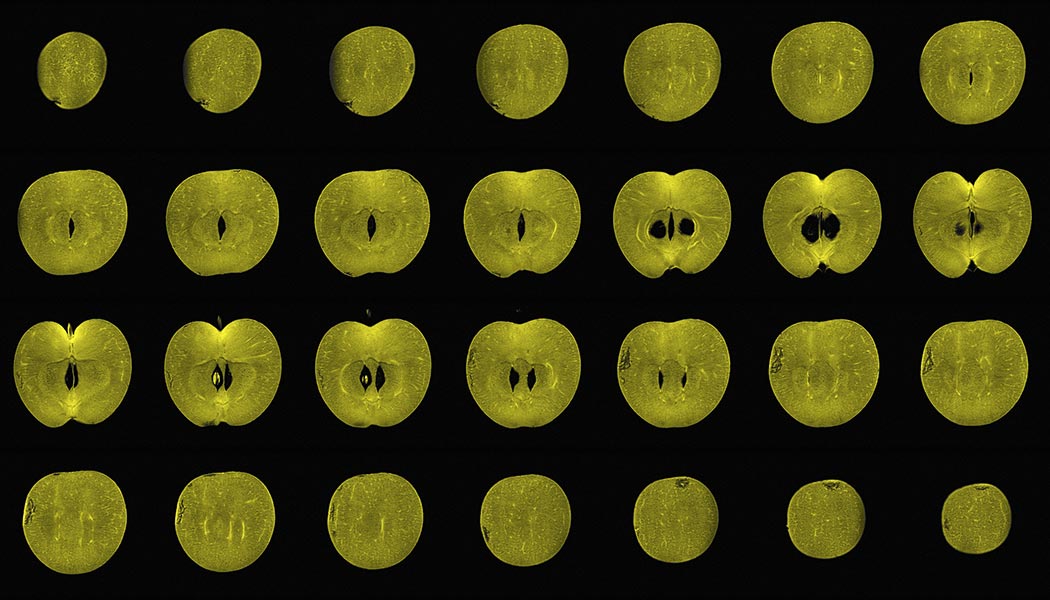
Below is an MRI image of a human skull. The second image shows an apple that has been virtually sliced down from the top to the bottom in a series of thin layers. These images were made by MRI which shows details inside the apple without damaging it. Image credit: Alexandr Khrapichev, University of Oxford. (CC BY 4.0)