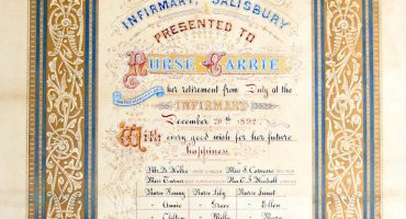
Memories of X-raying an Egyptian Mummy
We interviewed Radiographer Miles Woodford in November 2024 and in this extract we talk about his involvement in taking x-ray images of the Egyptian Mummy Hatemui. The conversation details the examination of a mummy using X-ray and tomography to determine the age based on bone development.
So we’re all behind the screens, and we do this, and we just drive this thing up and down, and there wasn’t a hair left bent, you know ‘dum! wow!’ you’ve got this wonderful, wonderful mummy. And the first time ever that anybody in, say, 3000 years, has actually seen what’s inside. It was just absolutely out of this world!
Listen to Miles talking about x-raying the Egyptian Mummy – MP3 file 6 minutes 8 seconds
Read the transcript of Miles talking about x-raying the Egyptian Mummy – PDF
Miles Woodford, was a senior radiographer at Salisbury Hospital, he discussed his career, starting in 1971 and spanning over 47 years, including his role in with the spinal unit in 1984. He detailed his travels to various spinal units in 1983 to learn advanced techniques, such as video fluoroscopy, which he introduced at Odstock. Woodford also described the evolution from film to digital imaging, highlighting the significant improvements in resolution and efficiency. He mentioned the historical use of X-rays for shoe fitting and the dangers of cumulative radiation exposure. The conversation also covered the shift from X-rays to MRI and CT scans for spinal imaging.
Read more about x-ray on our In your Bones project page