X-rays: the inside view
Our archive holds an exciting collection of old glass plate X-rays from the 1950s, as well as some earlier examples. Before we take a closer look we thought we should find out how these images help to diagnose illness and what an X-ray actually is.
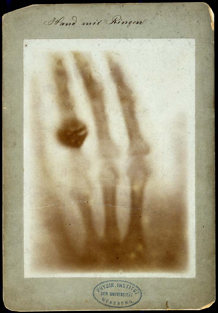
In 1895, the discovery of the X-ray was an important milestone in the history of medicine made by Wilhelm Röentgen, Professor of Physics in Bavaria. X-rays became more commonly used to treat soldiers fighting in World War 1, finding bone fractures and imbedded bullets. To highlight this look at the number of X-rays taken at Salisbury before and after the war. In 1913 Salisbury Infirmary took 248 X-rays but by 1919 they were doing 4 times this amount with 1073 X-rays.
What are X-rays?
X-rays are a form of electromagnetic radiation, similar to light. However, X-rays have higher energy and can pass through objects and in medicine they are used to make images of inside the body. Many of us will have had an X-ray, commonly at a dentist to look at teeth health or during an examination to check for broken bones. Did you know they were also used in shoe fitting up until the 1950s?
How X-rays work
- X-rays are a type of radiation that pass through the body. They can’t be seen and you can’t feel them.
- A detector on the other side of the body picks up the X-rays after they’ve passed through and turns them into an image.
- X-rays find it more difficult to pass through dense parts of your body, such as bone, and these show up as clear white areas. X-rays can pass through more easily softer parts, such as your heart and lungs, and these show up as darker areas on the images.
- Early X-rays, like those in our history collection were printed as a negative onto glass plates.
- Later X-rays were printed onto film and viewed on a light box. Today images are viewed as digital files on a computer screen.
Read more about how X-rays work
Glass plate X-rays – early examples from our archive
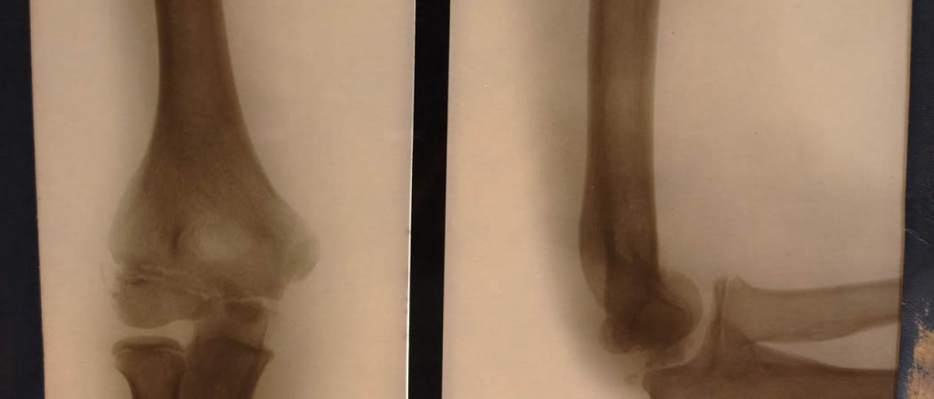
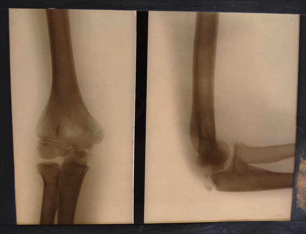
Large glass plate negative X-ray shows the elbow (humerous at the top leading down to the radius and ulna) is seen straight, left, and from front to back and with the elbow bent on the right.
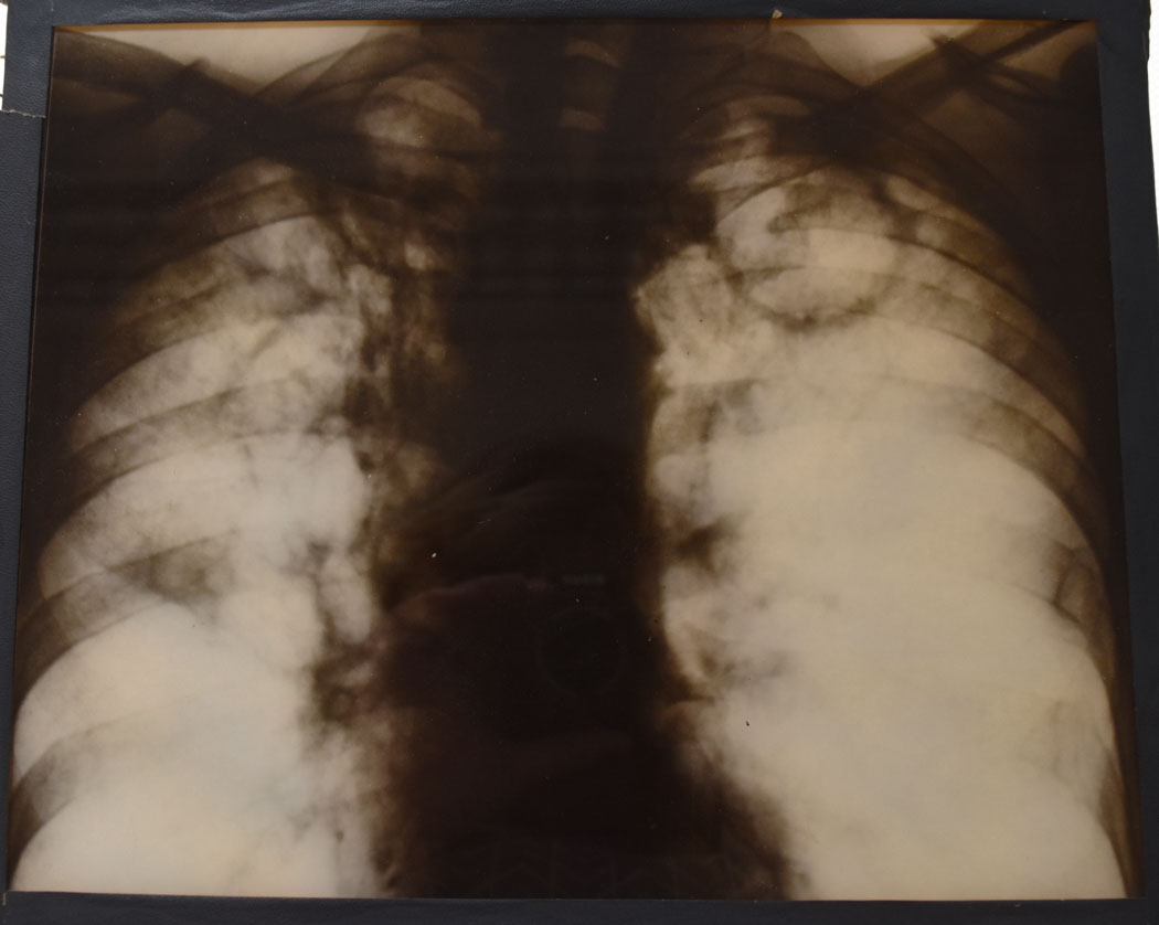
Glass plate image of a X-ray. This is a close up view of the upper part of the chest. The neck is at the top, then the clavicles either side and then the ribs. In the middle is the mediastum (trachea, main vessels and sturnum). Lungs are not clearly seen either side. Beginnings of the heart at the bottom of the plate.
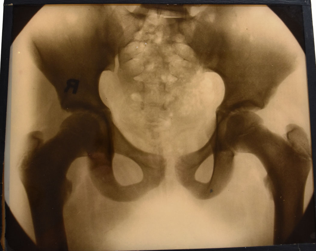
Glass plate image of an X-ray. The pelvis has the sacrum centrally and goes in a round and joins at the front. Joining either side are the hip joints with the femurs extending down. (Possibly female pelvis because of the wider pelvic cavity.)
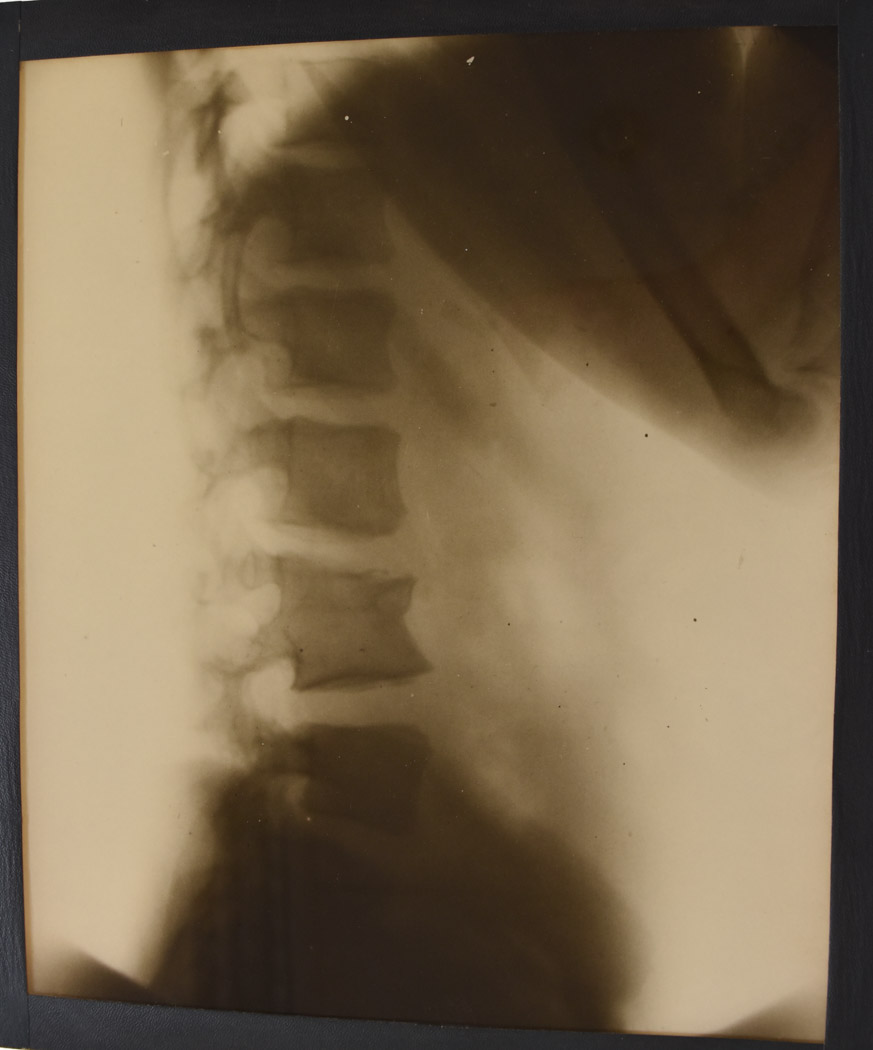
Glass plate image of an X-ray. Side view of the lumbar spine. The arms can be seen coming forward at the top with pelvis at the bottom. L4 (lumbar vertebrae of the spine) appears to have a wedge fracture.
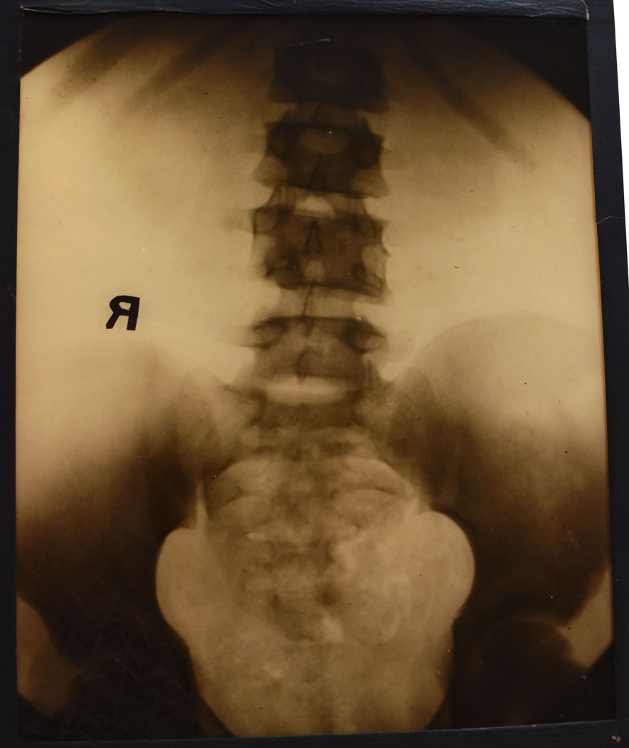
Glass plate image of a X-ray. The lumbar spine is seen centrally with lower ribs. At the bottom of the lumbar spine it joins to the sacrum , either side is the pelvis with the hips just coming off either side.
Glass plate X-rays – 1950s examples from our archive
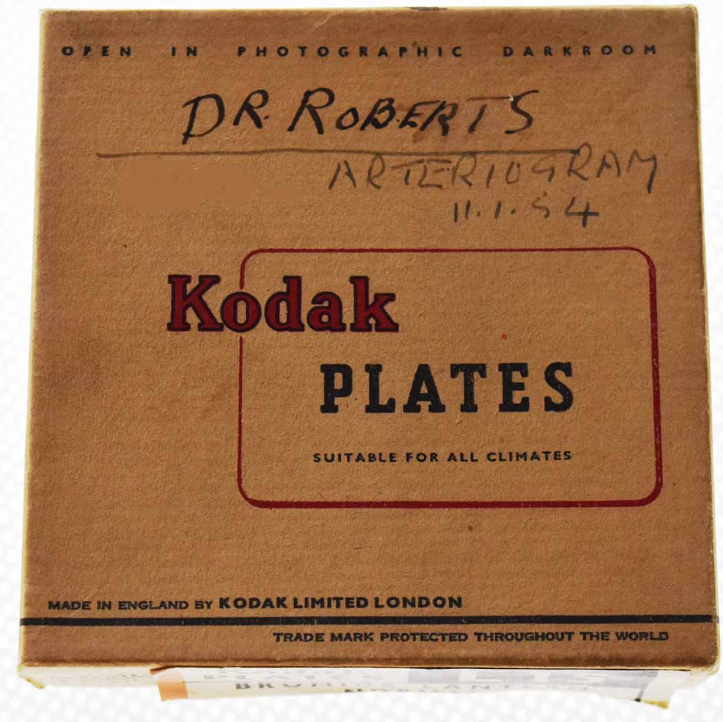
Cardboard Kodak box holding square glass plate X-ray marked Dr Roberts. It also says Arteriogram on the box and dated 11th January 1954.
Cardboard Ilford box for glass plate X-rays marked Dr Roberts. The box has information about how to develop the slides with the chemical materials that were used along with processing details. Dating from early to mid 1950s.

Collection of small square glass plate X-rays and microscopic slide sections, dated early to mid 1950s.
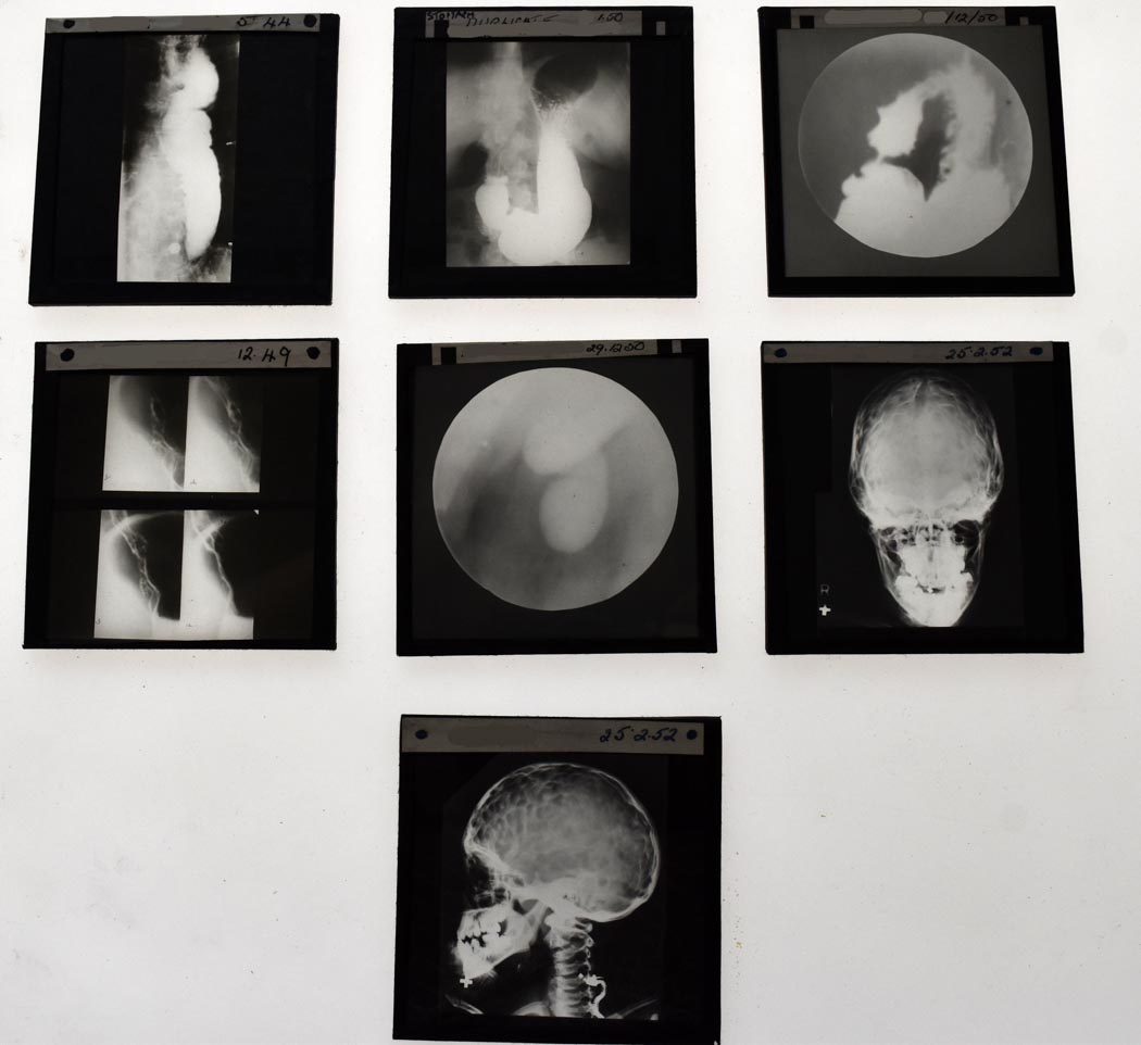
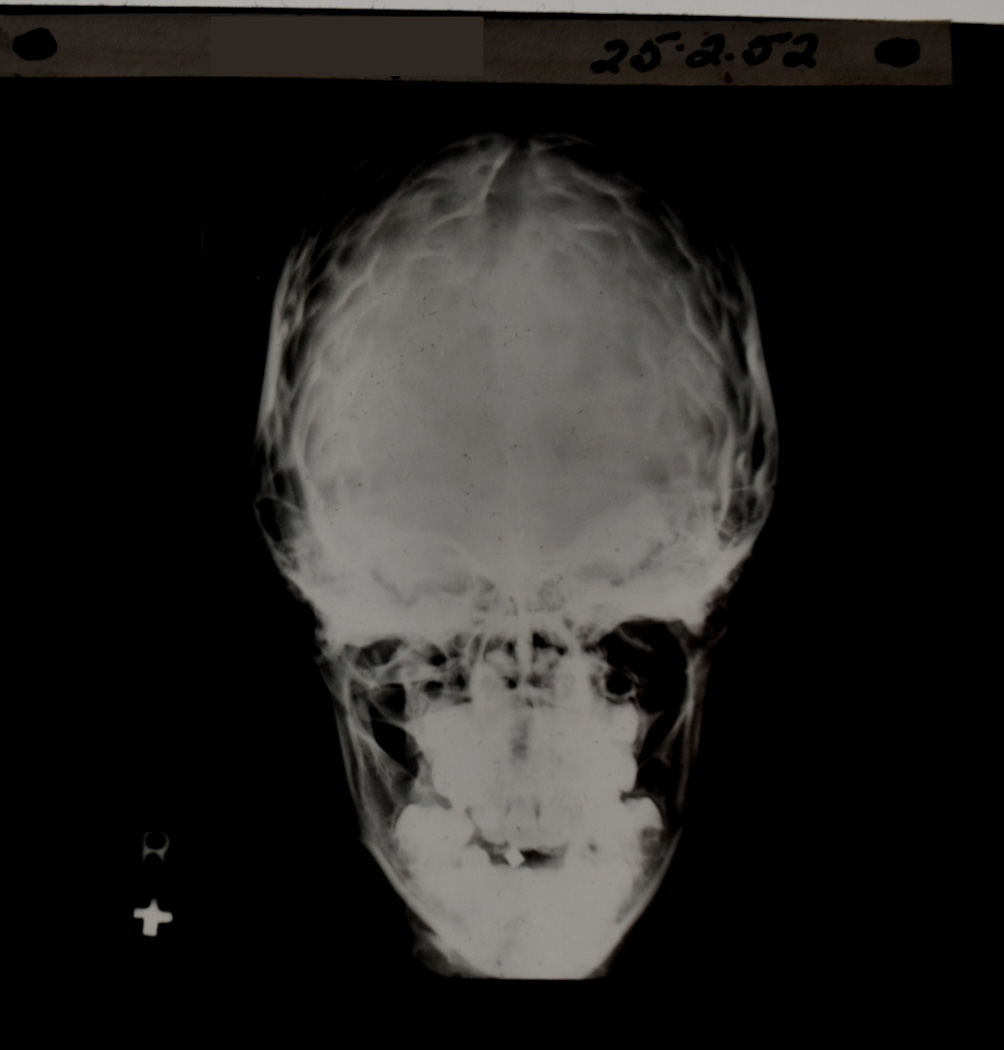
Square glass plate in a Ilford plates box marked Dr Roberts. This is a view of a skull looking from front to back . Main cranium at the top jaw at the bottom. Dated 25th February 1952.
Square glass plate in a Ilford plates box marked Dr Roberts.This is a view of a skull looking from the side, the neck is turned. At the bottom by the neck you can see a zip and a hand seems to be supporting the jaw. Dated 25th February 1952.

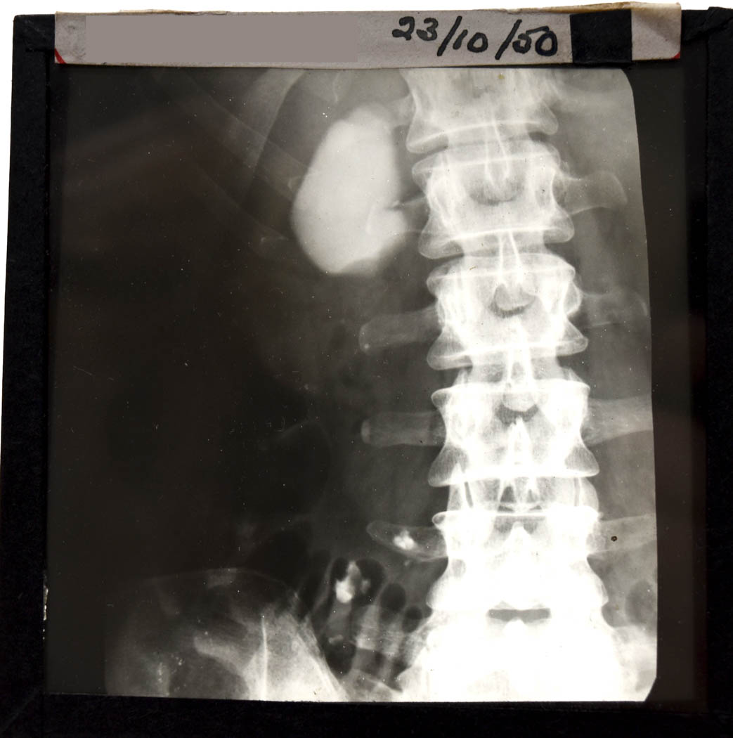
Square glass plate in a Ilford plates box marked Dr Roberts. The gall bladder is the bag shaped structure. Spine to the left and pelvis below. Dated 23rd October 1950.
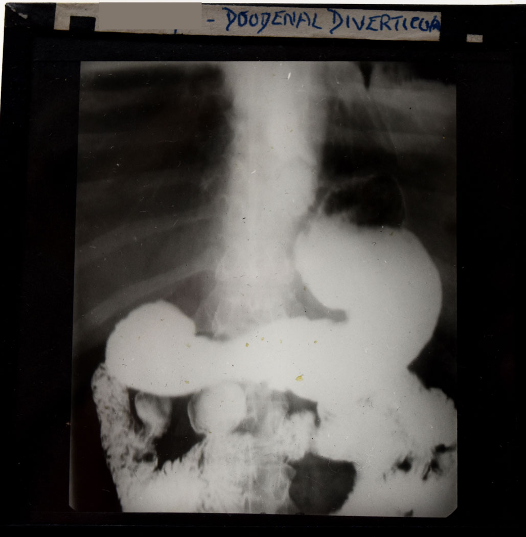
Square glass plate in a Ilford plates box marked Dr Roberts. View of a barium examination of the stomach and small intestine. The barium is the area in white. The spine at the back. Dating from early to mid 1950s. A barium meal test is used to see the oesophagus, stomach and duodenum. Because these softer tissues don’t show clearly on X-rays the patient swallows a barium meal which then shows up white on the images and reveals their shape. It helps to look for problems such as narrowing, hernias, tumours, reflux, and ulcers.
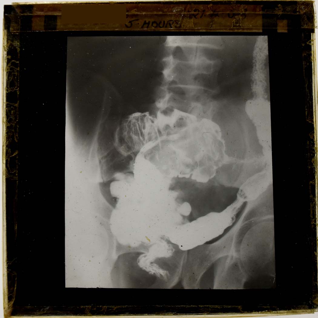
Square glass plate in a Kodak plates box marked Dr Roberts. This is a barium view of the bowel. The white is the barium the lower spine and pelvis is seen behind. Image taken 5 hours after start of examination. Dating from early to mid 1950s.
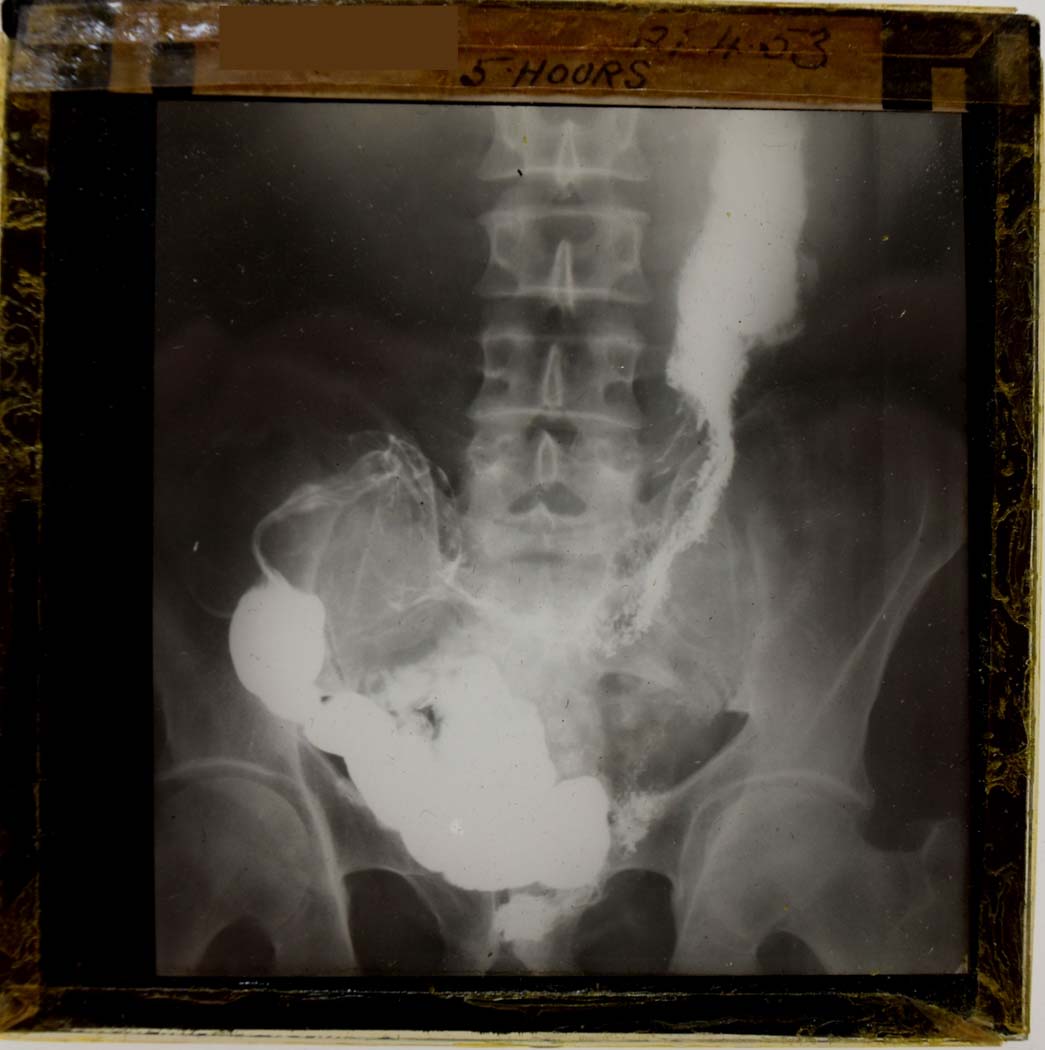
Square glass plate in a Kodak plates box marked Dr Roberts. This is a barium view of the bowel. The white is the barium the lower spine and pelvis is seen behind. Image taken 5 hours after start of examination. Dating from early to mid 1950s.
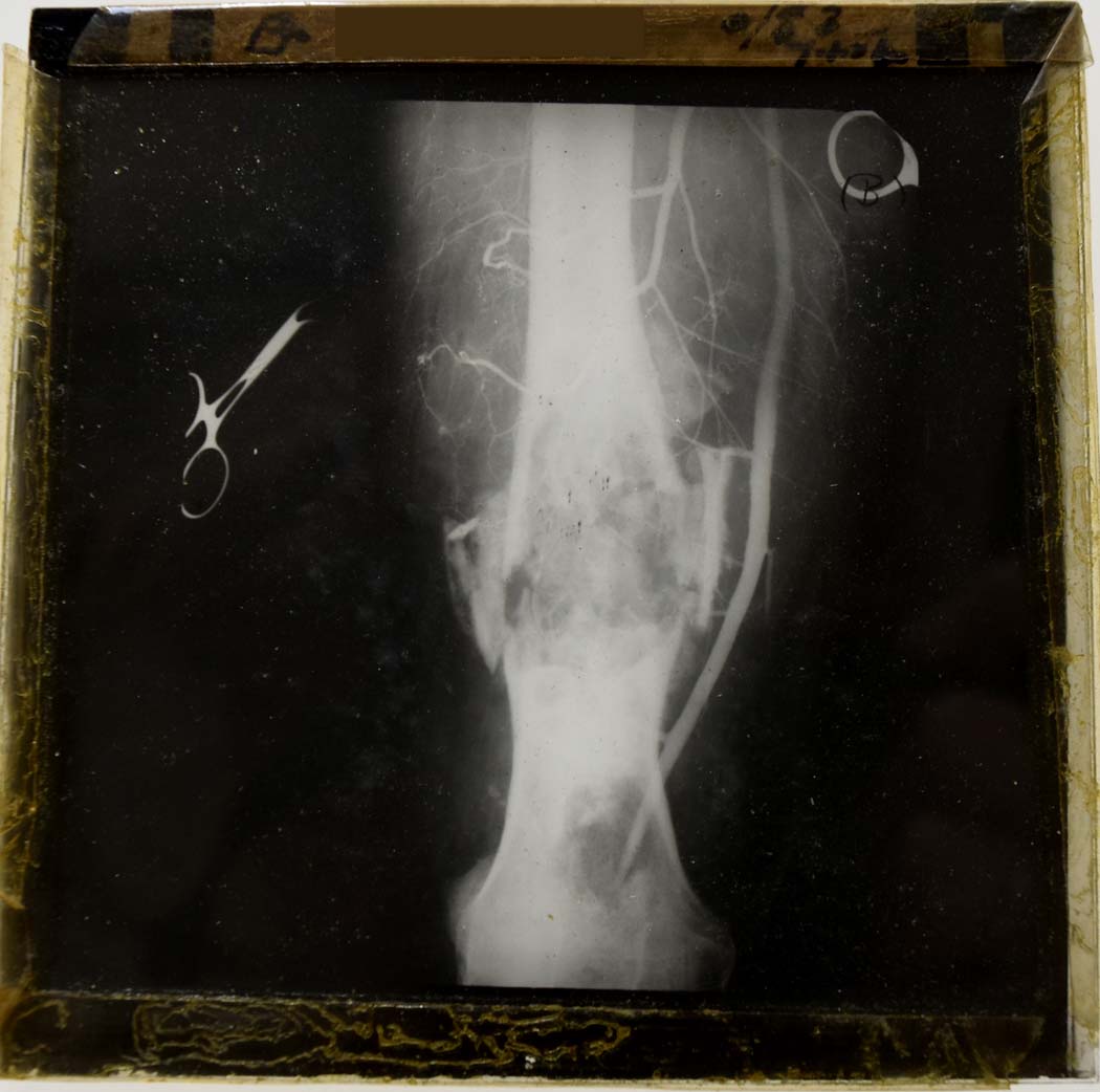
Square glass plate in a Ilford plates box marked Dr Roberts. View of a badly fractured femur, fragments of bone can be seen around the fracture the knee is at the bottom. The leg vessels can be seen in white around the femur due to contrast dye being injected. Forceps can be seen at the side. Dating from early to mid 1950s.
Read more
- All about X-rays – photographs and items from our collection showing the development of this technology
- X-ray shoe fitting – it was a thing!
- X-ray painting with light – try an X-ray themed creative activity